


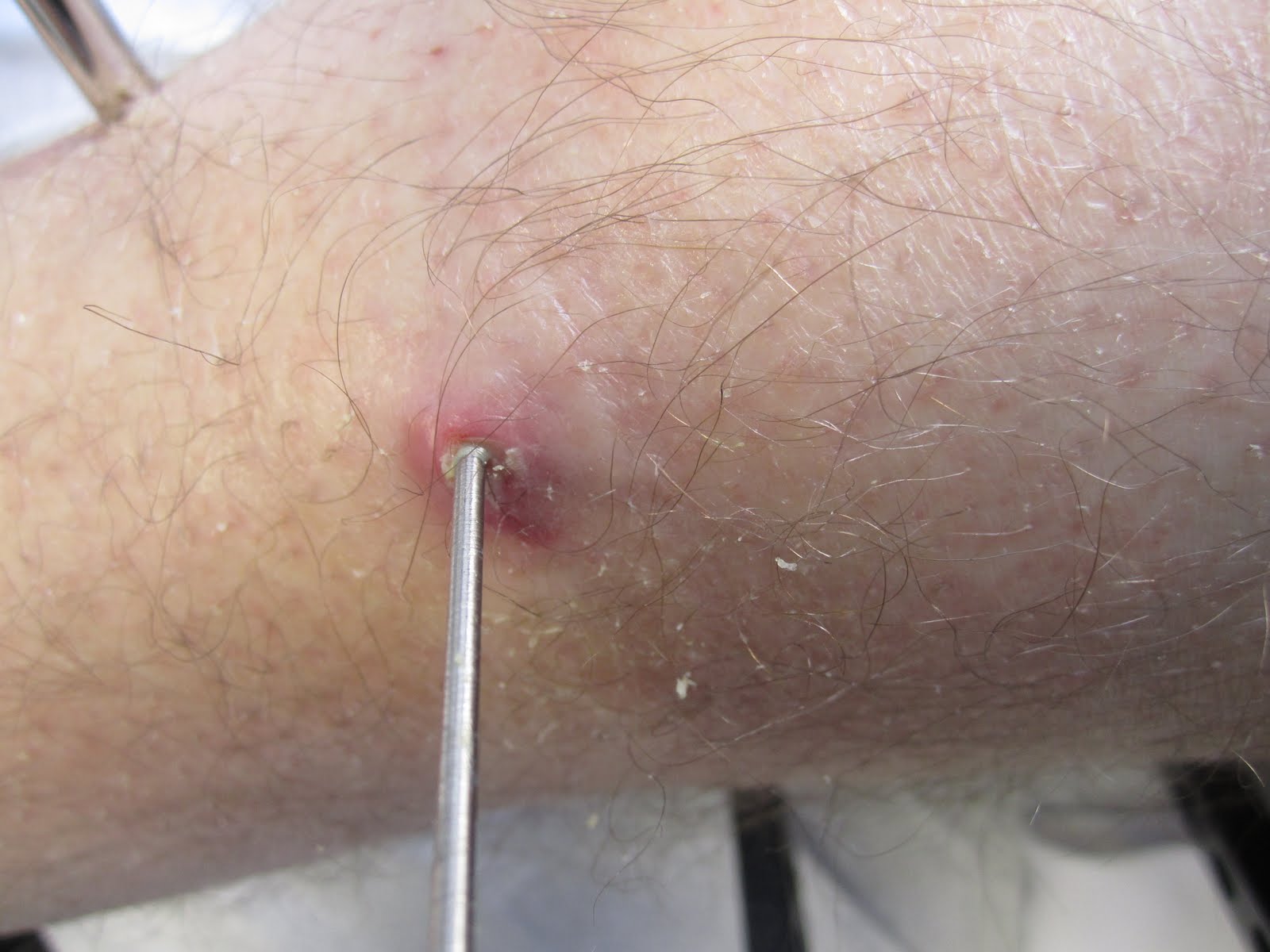


It looks like it's having a great time being infected.


Dr. Jeng was walking in and out of the room quite a bit today. Each time he came by, he'd bring in something new. Eventually he got his hands on the tools he needed from the operating room to remove the wire. There they are sitting on the table across the room, wrapped in blue paper.
I believe he had asked for a giant sterile wire cutter, a sterile 10mm wrench, and some pliers.

When Dr. Jeng came in for the last time, he took another look at the pin sites and the x-rays. The wheels in his head turned for a second and wondered aloud if it was worth taking the pin out this late in the game.
With each pin site removal, we inherit more risks with weight bearing, e.g., fractures in the bone, ex-fix breakage, etc. Bearing weight and walking around on this thing is absolutely critical to having any chance of a successful outcome. Because the x-rays did not show any problems, such as a fracture or an infection in the bone, Dr. Jeng reasoned that the infection in the pin sites was something we could control with antibiotics.
So at the last minute, Dr. Jeng called an audible and decided to put me back on antibiotics (Augmentin) to try and buy us some time before the November 3rd removal. I was on board with his change-up for two reasons:
- I want to continue bearing weight for the next three weeks to maximize the benefit of this procedure
- I'd love to avoid the pain (and crazy anxiety) of having the pin removed today
While this is probably the best decision going forward, I unfortunately continue to have pain that we've yet to get under control with any of our pain medications. After discussing this with Dr. Jeng and Brooke, we reasoned that the pain I'm experiencing now is a result of over doing it with unassisted weight bearing on the ex-fix.
It's true. Sunday night I was walking around the apartment without crutches like I was a 27-year-old with no right ankle arthritis. Starting the next day the unbearable pain came into the picture. Cause and effect.
Lesson learned: never do things you'll regret the next day.

On a good note, while we were looking at the x-rays, Dr. Jeng noted that the ankle distraction has noticeably improved my ankle arthritis. In the picture above is a six week comparison of my ankle with the most recent image from today on the left.
The first thing you might notice is the difference in joint space. Today I was not bearing weight and six weeks ago I was. Nothing to discuss here.
Another thing you'll notice is the difference in brightness of the bone near the joint. When bone shows up bright white on x-rays, that typically indicates hard, dense, unhealthy bone. This is a result of the two bones grinding against one another and causing the subchondral bone at the surfaces to harden -- particularly with so much cartilage loss. This is one source of ankle arthritis pain.
The bottom line is that the ankle distraction has allowed the bone to heal. The cysts and lesions are going away and the bone is not as bright, suggesting that the subchondral bone at the surface is returning to a more normal and healthy density level.
All awesome news. I'll continue to rest the ankle until some of the pain is under control and try to monitor how much walking I do on the ex-fix when I'm feeling good. Only three more weeks until we get to test out the joint after a twelve week distraction.
One last thing we asked for before we left today was the post-op surgery report. It was very interesting and went over my head several times. I tried my best to look up some of the medical phrases and terms and link those in so the report is easier to understand.
Procedures Performed:
- Right ankle arthroscopy and debridement, anterior cheilectomy
- Distraction arthroplasty and application of multiplanar external fixator
- Tendo Achilles lengthening
Anesthesia:
- General, popliteal block and saphenous nerve block
Complications:
- None
Tourniquet Time:
- 120 minutes at 350 mmHg
Implants:
- Smith & Nephew Taylor spatial frame, four olive wires [I've been calling them Kirschner wires; I've yet to figure out the difference], two smooth pins, two hydroxyapatite coated half pins, and then a standard Ilizarov Smith & Nephew frame with two Taylor spatial frame hinges and two universal hinge struts.
Findings
Mr. Meehan is an amazing, handsome, extremely intelligent, and gifted athletic 29-year-old gentleman [I added the adjectives.] that had post-traumatic injury to his distal tibia and has undergone multiple prior surgeries first for open reduction and internal fixation and then subsequently sustained an infection to his ankle joint [This part is not exactly correct] which was washed out and then subsequently had his hardware removed. However, in this time he has developed degenerative joint disease in his right ankle with significant arthritis and osteophytes and subchondral cysts seen on his plain films and his CT scan. He has significant pain with ambulation and has seen various physicians for surgical options with regards to his right ankle. He was given options previously of fusion versus distraction arthroplasty and does not wish to proceed with any type of arthrodesis of his ankle. He has had no evidence of any infection in his ankle since his last wash-out [I've never had a wash-out surgery]. There has been no drainage. It was discussed with him various options including nonoperative management, arthrodesis versus distraction arthroplasty and given the patient's desires, he wished to proceed with the distraction arthroplasty. It was discussed with him that this may not be successful in the short-term but may be successful in the long-term or with a five-year period. The risks of the surgery included but were not limited to bleeding, infection, pin track infection, persistent pain, scar, stiffness, incomplete relief of preoperative ankle pain, hardware breakage, need for future revision or reconstructive surgeries, reaction to anesthesia, stroke, heart attack, loss of limb or death. The patient understood all the above risks and wished to proceed with the surgery [I probably didn't understand all of the above risks]. All his questions were answered in detail and informed consent was obtained from him. A copy of the consent form was signed by the patient and the attending physician and appropriate witness. A copy of this was placed in his permanent medical record [I didn't realize permanent records existed].
Description of Procedure
On the afternoon of 8/15/11 the patient was taken to the Mercy operating room and laid supine on the operating room table. He underwent general anesthesia via laryngeal mask airway and received 2 gram of Ancef intravenously. At this point a tourniquet was applied to the right upper thigh and then a timeout was called to correctly identify the patient, operation, operative site and all in attendance were in agreement and the procedure was begun, again by starting with a popliteal block, using a nerve stimulator with 40 ml of 1% Lidocane and 0.5% Ropivacaine. After this was performed the right lower extremity was prepped and draped in the usual sterile fashion. A tendo Achilles lengthening was made with three percutaneous stab incisions, two medial and one lateral given that the heel was in slight amount of valgus. After this was done the leg was elevated and the tourniquet was inflated to 350 mmHg. We first identified the anteromedial entry point with normal saline and then insufflated the joint and then placed the heel distractor on the patient and then distracted the ankle.

Heel Distractor
We made our anteromedial portal with a nick and spread technique and then inserted the arthroscope and then began our diagnostic arthoscopy. There were significant findings for complete cartilage loss on both the talus and the tibial plafond consistent with his preoperative radiographic imaging. There were very small islands of cartilage medially on the talar head [This explains why most of my ankle pain was on the lateral side], however, there was significant degeneration widespread throughout the joint. There was extensive synovitis which was visualized [This explains a lot of my pain]. We made our anterolateral portal under direct visualization with needle localization and then using a nick and spread technique inserted the shaver through the anterolateral portal [The camera was inserted on the medial side and the shaver on the lateral side] and debrided the extensive synovitis and as well as removed the anterior bone spur on the tibial plafond, performing and anterior cheilectomy. The patient again had very little cartilage on the talus or the tibial plafond remaining, but given the extensive nature of the arthritis, there is no area to specifically respond to with the microfracture so thus after doing our debridement cheilectomy, withdrew the instruments and then took traction off of his leg.
Following the ankle arthroscopy being completed, we then turned our attention toward placement of our Ilizarov frame. We used a standard Smith & Nephew two ring foot frame. We slid this over the foot and ankle onto the tibia and then identified the tibial crest and then placed a half pin through the most proximal frame, which was hydroxyapatite coated. Following this a second half pin was inserted along the medial face of the tibia for the second ring.
These were fixed to the frame and the smooth wires were then drilled in a perpendicular fashion to these half pins and then one was attached to the proximal ring and another to the distal ring and then these were both tensioned to 130 pounds per square inch and then at this point, held in place through the Russian smooth wire holders, the appropriate nuts were tightened to hold the wires in place. After this was done we then placed two olive wires in a cross fashion into the calcaneus [heel bone], again tensioning these to 90 pounds per square inch, and then sequentially tightening these through the Russian clips and tightening these with the nuts, and finally two more olive wires were placed in a cross fashion through the forefoot obtaining purchase in the fourth and first metatarsals, avoiding the fifth metaphalangeal joint [my pinky toe joint] and were tightened in a similar fashion, tightening to 50 pounds per square inch. At this point manual distraction was placed on the ankle and then using the four struts around the frame, the distraction was held in place and locked and then using fluoroscopy we checked the amount of distraction on both the anteroposterior and lateral views. We noted the cheilectomy that we had performed had smoothed out the anterior aspect of the joint and that we had obtained an increased amount of ankle distraction. These were then held in place and then the malleolar axis was identified under fluoroscopy, drilling from medial to lateral, from the tip of the medial malleolus to the lateral malleolus [they drilled in a pin through my ankle joint to line up the two universal hinges on the back of the ex-fix]. Using this as a guide we then placed universal hinges along the axis of the ankle, one medially and one laterally, and then locked these in place and then further distracted the ankle and locked these in place. Following this the four prior struts that were used were removed and loosened. We noted at this point that we could get 10 degrees of dorsiflexion and 20 degrees of plantar flexion of the ankle on the table. The two Taylor spatial frame struts were then applied to the anterior aspect of the ankle and then these struts were locked in place. Following this all of the half pins were cut and then the foot plate was attached to the bottom of the frame. Please note that all of the sharp ends of the half pins were capped and then the cut edges of the smooth pins and olive wires were bent and curled into the anterior aspect of the frame. The wounds were then cleaned and dried. The arthroscopy portals were closed with a 3.0 nylon and then covered with Xeroform and then 4 x 4's and over-wrapped with Webril. The pin sites had sterile gauze applied over them and then had foam spacers placed along them toward the outside of the frame and the frame was over-wrapped with an Ace bandage and then the patient received a proximal saphenous nerve block for residual postoperative pain relief.
At this point the patient was awoken from his general anesthesia and then transferred to the recovery room in stable condition. At the end of the case all needle, sponge, and lab counts were correct. Dr. Jeng was present and participated in all aspects of the surgical case.
I knew it...prayer works!! So glad to hear the news, although I have to be honest, Josh is really pullin' for the bionic leg option. We love you and will keep praying. Three more weeks, no problem!! Rach
ReplyDeleteHi, thanks for your posts. Good luck, I'm looking forward to hearing your results. End-stage ankle rheumatoid arthritis at 48 y/o for me. Not liking the options of fusion or replacement so I'm hoping that extraction continues to prove promising. Thanks also for literature review. Be well!
ReplyDeleteJB
JB - Sorry to hear about the rheumatoid arthritis. Before my accident, I had no idea how debilitating arthritis could be.
ReplyDeleteI'm working on a summary of the research articles on ankle distraction, so stay tuned for that. Mine will be focused on osteoarthritis, but I've got some good data on the procedure and outcomes for ankle distraction arthroplasty.
Man! You cannot imagine how you helped me with this blog! Last July I had a freak accident (not as glamorous as yours, only fell from a ladder) and busted my right angle pretty bad. Went throu similar things (emergency surgery, later infection, onset of post-traumatic arthritis), and now this uncertain road between ankle fusion, replacement, etc. Last month I felt really really depreassed, to the point my wife noticed it, but then I found your blog with your attitude and approach to the issue, really inspiring! Thanks for that! In the meantime we are here in the south putting the same battle. Wish you the best and thanks again!
ReplyDelete