Everyone has a Mom. As you know, a Mom's primary job is to be concerned.
Tomorrow at 11:00 AM I'll be getting at least one Kirschner wire holding the most proximal Ilizarov external fixator ring to my tibia removed. As Dr. Jeng put it, he wants to get rid of my "soupy pin."
The plan is to get fresh x-rays of the pin sites to see if there are any issues with the tibia bone and the pins. If the images reveal any issues, there's a chance we'll remove more k-wires or just take the whole apparatus off since we're close to having this thing on for twelve weeks.
This is Step One to freaking out my Mom. She's far away and won't know what's happening until after it's already happened. In other words, she's worried about not being able to worry about what's specifically happening to me.
Step Two is showing my Mom new developments with my ankle without first running it by her on the phone.
Last night, Brooke and I discovered something interesting I can do with my new Achilles tendon. During my August surgery, my Achilles tendon was lengthened with three cuts. These cuts left three dark marks along the tendon.
When I flex my calf muscle, it appears that the skin, muscle, and fat all pull along the sites where the tendon was cut. In other words, I've now got awesome calf muscle definition that most people dream of. My definition just so happens to be severely deformed.
We'll talk to Dr. Jeng about what's going on here tomorrow, but I suspect it's scar tissue that's formed around the incisions made in August to lengthen the tendon. With physical therapy, this odd deformity will probably go away. This deformity might also be due to the fact that the joint is pulled apart by 5mm. This might also go away once the ankle is no longer distracted.
Step Three is to show more cross leaking pictures, including a new leaking pin site, and note that pain has become an increasing issue in the last few days.
This is the original soupy pin that will be removed tomorrow. Nothing new here -- it's still leaking.
Unfortunately, the lateral side of the second-most proximal k-wire is also leaking now. There's a chance that both of these k-wires get removed tomorrow.
But if those two wires are removed, then the argument for keeping the external fixator on the leg becomes harder to defend -- particularly with November 3rd just around the corner.
We'll let you all -- especially you, Mom -- know how it goes tomorrow!
Let me quickly tell you how this post is going to go. It's going to start out gross, get grosser if you watch the video, then get sweet and sappy.
That's just how I do it, especially when the Redskins are down to the Eagles. At least the Redskins have realized it's time to pull Grossman and put in Beck.
Over the last week or so, I've experienced more pain and soreness in all fourteen pin sites -- particularly the proximal k-wire on the lateral side of my leg and the two pins on the lateral side of my fore foot. In order to sleep, we've had to resort to some of the heavy duty medication from several weeks ago to keep the pain managed.
As a quick aside, walking on an external fixator is a totally different experience than passively wearing one for three months. Last year, I was off pain medication within two weeks of the surgery. This year, it seems like as time goes on, pain medication is becoming more important to help me tolerate bearing weight. Bearing weight is an essential part of ankle distraction arthroplasty, as I'm learning from the research, so it's extremely important to me to continue walking on the ankle.
Part of my problem is edema. As the ankle and leg swells with fluid, extra pressure is exerted on all of my pin sites. If the pin site is not sealed, it will spring a leak (so to speak). The edema fluid will dry around the pin site and form a crust -- kind of like a scab. As the edema continues to build up, the skin inflates around the dried crust and causes irritation.
It's an endless cycle of awesome.
This is the proximal k-wire pin site on the lateral (outside) side of my leg. This picture was taken yesterday. Of all of my pin sites, this guy has caused us the most grief since mid-September. While edema is a problem here, Dr. Jeng and I are more concerned that either a latent infection is causing some issues or, worse, a partial fracture around the bone might be allowing the k-wire to move as I bear weight, irritating the muscle, bone, and skin when I walk.
The fluid leaking out of the site does not appear to be infected. It's clear and yellow, indicating that it's edema fluid. However, the pin site itself is continuing to get more red and the fluid is now draining from the site like its a bathroom faucet.
Warning: this video is gross.
You'll notice two things in this video, taken yesterday. First, the lemonade is indeed flowing from the pin site like a bathroom faucet. Second -- and this one takes a keen ear -- you can hear a clicking noise from my shin muscle rubbing up against something -- either the k-wire or the half screw.
After sending this video and other pictures, linked above, to Dr. Jeng last night, I got an email from him this morning:
We should get the soupy pin out this week. How many more weeks until 12 weeks?
Hence the Campbell's Wire Noodle Soup picture above. So it looks like on Tuesday we'll be taking this k-wire out. My immediate concern is whether or not I'll be allowed to bear weight without this k-wire. I assume he was asking me for the removal date to help him decide whether or not I could continue bearing weight.
We're currently sitting at three weeks until I've worn this ex-fix for twelve weeks. We'll see what happens Tuesday. I imagine we'll also take some X-Rays of the mid-section of my tibia to look for hairline fractures near any of the other pin sites. I will, of course, have video of the k-wire removal.
Now that the gross part is out of the way, now it's on to to the sappy and sweet part of the post. A few months ago, I was joking with my Grandmother that she and I should get matching canes. We were discussing with one another that we needed to setup an official race to see who was faster on their feet.
She was challenging me to a foot race. And, worse, I was very worried that she'd dominate.
Last week I had two custom canes made for both of us. It's a golden sienna derby walking cane with a black beechwood shaft and silver collar. In other words, it's extremely fancy.
More importantly, both of our canes have a custom oval engraving that includes our initials and our position in the Foot Race Society. My Grandma ended up beating me in the presidential race, so she's the President of the Foot Race Society and I'm the Vice President.
I'm hoping this stops her from continuing to challenge me to a foot race. Like I said, I'm fairly certain she'd beat me every time.
The handle is my favorite part. I know you're jealous. You want one, I know.
Here's the President of the Foot Race Society proudly displaying her new can along with her beautiful October decorations outside her country home.
And here is the Vice President, proudly displaying the exact same cane in front of a deadbeat dog in the background that only knows how to sleep, eat, and fart.
I'm forcing a smile here because I just found out the Redskins lost.
In March 2010, just a month after my accident, I was curious to see how much it costs to "fix," my ankle. I put fix in quotes because, obviously, we're still in the fixing stage. I titled that post, "American Healthcare."
It's been a hot topic in our country for a while.
After adding up the costs, my first reaction was shock. Over the three days I stayed at the hospital in Colorado, my medical bills exceeded $73,000! I found out later from a friend of ours that operating rooms bill you and your insurance company the same way Verizon and T-Mobile do it: by the minute.
That seemed nuts, so I Googled. According to this Consumer Reports article from 2009, "operating-room use is generally billed at rates that vary from $69 to $270 per minute."
That is nuts.
My second reaction at the time (and still to this day) was relief at how lucky my wife and I are to have jobs and to be covered by a good health insurance plan. Not once have we had to argue or dispute anything. Moreover, they've covered almost all of my costs.
Today I took a quick look at what providers have billed my insurance company since my accident on 13 February 2010 to 25 September 2011 (so we're still missing several visits to my family physician and surgeon).
The grand total: $288,947.54.
My insurance company negotiated those bills down to $161,986.16, a difference of $126,961.38 (a 44% discount).
Our out-of-pocket expenses so far have been $6,950.81. That's 2.4% of what we've been billed and 4.3% of what my insurance company negotiated with each provider. Another important thing to note is this out-of-pocket figure does not account for the hundreds, if not thousands of dollars we've spent on our own to help manage wounds, the ex-fix, and other rehab equipment. I'm betting our out-of-pocket figure is probably over $9,000.
The crazy part about all of this is we are not even close to being done with my ankle. My guess is we'll reach half a million before all is said and done.
On October 2nd, I wrote a post about how to prepare for your first external fixator. One of the items I recommended was a new set of replacement crutch tips for the low, low price of $8.99.
I received my new extra large crutch tips on Wednesday, the 5th. Eight days later, the replacement tips were dangerously unusable.
Please keep in mind that I don't do much on my crutches. The most activity I see in a day is racing for the remote control on the other side of the couch when Grey's Anatomy comes on.
I have to make sure the volume is up high enough for me to follow TV's most compelling prime time drama.
I had two problems on each tip. The first problem was a minor tear in the grip. While this isn't terrible, these kinds of tears make it more important to keep your center of gravity over the points where your crutch tip meets the ground.
I'm not sure how I got this tear so quickly. I crutch on carpet, tile, hardwood floors, and rarely, sidewalks.
Here's problem number two. This is the most dangerous type of crutch tip break down. It highlights the importance of examining your crutches every morning and evening.
When the crutch tube is poking through the tip, you've lost all grip. At any moment, you could place too much faith in the gripping ability of your tips and end up having the crutch slip out from underneath you.
You could fall on your ex-fix or, hurt your other ankle, or even worse, injure your dignity.
Do not buy these replacement tips. They are terrible. I should have read the review, "Do Not Buy - They wore out in one week," before buying these tips. That guy was right. All it took was one week.
Instead of getting these replacement tips, ask your friends for piggyback rides.
This is my good friend Alison giving me, a 6'3" 215 lbs. (all lean muscle) man a piggy back ride on a bet that she couldn't carry me 30 feet. This is the same Alison that won the inaugural Injured Tony Award. To Alison's credit, she dominated it and carried me the entire distance.
The ironic and funny part of the story -- Alison injured her ankle.
Oh, no. Stop right there. She didn't injure it while giving me a piggyback ride. That would make sense.
No, she injured her ankle after trying to give me a high five to celebrate her performance. She had a poor landing on the way down from slapping skin. Perhaps I held my hand too high, or maybe she tweaked something during the piggybacking.
Whatever the reason, the lesson of this story is very clear: Alison is terrible at high fives, so don't buy these replacement crutch tips.
Have you seen the movie, Groundhog Day? If you haven't, leave right now, run to your nearest Blockbuster, grab a VHS or DVD (if you're lucky, Blockbuster should have one last rental sitting on the shelf in the Awesome Movies aisle), and park yourself on the couch for one of 1993's best comedies.
The premise is simple: a weatherman, played by Bill Murray, finds himself living the same day over and over again.
In many ways, the last two years of my life are taken right out of Groundhog Day. Instead of reliving the same day, I'm stuck in the same year -- over, and over, and over.
As the movie teaches us, reliving this experience is an opportunity -- a gift to learn how to be a better man. You'd think I'd learn. You'd think I'd be a able to handle the daily onslaught of questions from strangers about my ankle a little better.
I'm afraid I've learned nothing.
I usually stick with, "it's a snowboarding accident."
But some people are smart and quickly realize that it's not winter yet. So the questions continue. I average about ten new strangers a day. Each encounter is about a minute. That's over an hour a week of me talking about my stupid ankle.
Look -- I don't mind taking time to tell people how I was an idiot and snowboarded into a tree two years ago. I just occasionally wish these people would organize, form a board and write a charter, and then ask me all of their questions. Their secretary could write it all down and we'd be done. I'd just direct all of the new strangers to the board and have them review the minutes from previous meetings.
My friends have also suggested I get a business card with a link to this blog. That's probably a more practical idea. And not dumb.
An Idiot's Guid to Snowboarding into Trees
http://snowboardervstree.blogspot.com
I'm always polite and I try to keep it humorous to put the other person at ease. I really want people to feel comfortable and feel free to ask anything. Usually, the conversation starts because the other person is genuinely concerned about my well being. But once the friendly stranger comes to the realization that, yes, the pins and screws are going all the way through the skin, muscle, and bone, things turn and the friendly stranger ends up needing the counseling.
Surprisingly, to this day, The Bear Trap post is the most visited on my blog. Is this because there are a lot of hunters (I'm assuming they're mostly Canadian) Googling for bear traps but are stumbling across a ridiculous post on the Internet?
No one knows. Probably.
Because I entitled this post, "Groundhog Day," I imagine I'll get a lot of hits from people either looking for the movie or what the holiday is about.
Sorry, dudes -- you're not going to get the answers you were Googling. Learn how to Google. Instead, you're going to get my 2011 list of alternative stories I need to start feeding to people.
10. I Didn't Make the Jump
First of all, from the very moment you hit play, it's obvious I'm not going to make it over the ramp. We didn't plan for enough road to allow my bike to get up to the 88mph needed to successfully clear the ramp.
Second of all, once I get to the edge of the ramp, I know the show's over. My brain should be coordinating a complex series of maneuvers to get me a quick, graceful, and pain-free exit out of the fall.
But instead, the brain uses that jerk tone of voice with me and says, "you got yourself into this, so I hope your face enjoys cement."
Then somehow I severely break my ankle.
9. I Watched an Intense Episode of House
I broke my left ankle just by looking at this creepy picture of House.
8. The University of Stanford Mascot
There are a couple of options here. The obvious one is I broke my ankle when I learned that one of the most prestigious universities in America has a tree as it's mascot.
Another idea is the Tree kicked my butt for making fun of how it routinely shows up in top ten lists for the worst college mascots in America.
Regrettably, I have little room to talk. This is my school's current mascot.
7. Troy and his Lu Lu Lemons
The other day, I was hanging out with my friend, Troy. He was wearing -- what I thought -- were regular eBay or hand-me-down sweat pants. It was a casual Sunday, and I wear either gym shorts or break-away pants all the time.
I'm not one to judge.
But then I found out they were Lu Lu Lemon pants. That changed the whole story. I did some judging and ended up breaking my ankle.
This one needs some work. I don't want to imply that Troy broke my ankle. That wouldn't happen.
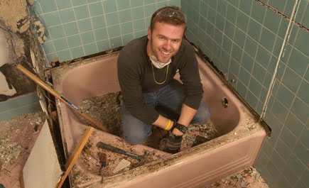
6. Bath Crashers
Have you ever seen this show on HGTV? It's freaking awesome! This guy hangs out at Home Depot all day asking random people if he can use their bathroom. Then he surprises you by tearing your bathroom apart and building your dream office-at-home.
I'm not sure how this breaks my ankle. I just think this show rocks.
5. Marital Argument
I lose every time. If I attempt at defending myself over why I left an empty bowl of cereal out in the living room, especially after my wife gets home from a twelve hour shift that ran fourteen hours long, bones get broken.
I'm not even making this one up. She leaves the bruises in places where people won't see them.
She doesn't read this blog. Help me.
4. Complainers
We all work and know people that incessantly whine and complain about everything -- especially little, insignificant things. If you've been around someone like this long enough, you know there's a good chance you're ankle will snap when you slam your foot up his -- sorry, I forget sometimes I've got family reading my blog.
It's easy to break your ankle around a complainer.
Also, if you don't know of anyone that acts like this, then you're probably the whiner in your group of friends.
Stop whining.
When people try to complain to me about a bruise they have on their thigh or a sore wrist, I wonder if they're color blind. And by color blind, I mean I wonder if they're incapable of seeing eight steel pins stuck in my leg surrounded by a bird cage.
3. Las Vegas with my Friends
This is a friend of mine getting a wake-up call from two of my other friends in Las Vegas this summer. And with the way my friends operate, it's a never ending war of escalating pranks.
We've already passed buckets of water. Broken ankles are only a few more pranks away.
2. Rock, Paper, Scissors Accident
First of all, yes, this is real. I haven't quite figured out yet how to break my ankle doing this, but I'm sure if someone threw rock hard enough, it could cause some problems.
Did you know that there's an actual strategy to this game?
Here it is. Now you know how to resolve all arguments by proposing (and subsequently dominating) a rock, paper, scissors game.
1. Air Guitar Melted My Face (and Ankle)
This is also real. It also sounds just as stupid as a rock, paper, scissors league. Back in 2009, a few of us decided to go watch a live regional competition in Washington, D.C. We thought it would be terrible.
It turned out to be AWESOME.
This is C-Diddy. He's retired. But this is what started it all. Some of these guys are so good, they can easily break ankles.
Summary
That's my list. Let's see if we can outdo The Bear Trap post. I'll have some updates this weekend of a sweet new custom cane I had made for my Grandmother and myself. You'll be jealous (for about a second or so) that we get to use canes and you do not.
Across the world today, several of our close friends ran a marathon -- two for their first time.
Congratulations to Katy (left) for completing her very first marathon in Chicago with seasoned marathon runner Crystal. We're very proud of both of you! Nice work on a personal best time, Katy!
Halfway across the world in Oslo, Norway, our friend Ken also finished his first marathon. We're not as proud of him, though.
Why?
Hopefully this picture of Ken should explain why we do not care. Also, as a side note, if you're familiar with the Internet, you might recognize the following picture and see a close resemblance to Ken.
Days like today I usually make fun of my friends for having to work (or, apparently, run for 26.2 miles) while I sit on my couch and watch Sports Center all day long.
It sounds good on paper. I always enjoy making fun of my friends. But there are only so many times I can watch an ESPN segment about whether or not Tim Tebow should get the start against Miami or see what LeBron James had to say about it on Twitter.
Of course Tebow should start. Quit arguing about it every 45 minutes like there's a choice.
The ex-fix feels great. I'm able to walk around the apartment without crutches and it feels comfortable. Of course, after walking around like that for a few minutes, the pin sites get sore and Percocet is required to quiet down the pin site pain.
So I guess I shouldn't say it feels great. I should probably say it feels great until I use it. Then it feels terrible.
The proximal pin site on the lateral side, the one that's been giving us some grief for some time now, still won't quiet down and scab up like the rest of the pin sites. After walking around I usually start bleeding out of the site (as seen above).
This picture was unusual because the blood seeped through the Xeroform wrap we placed around the pin site. We would have expected it to hold in some of the drainage.
We decided to employ some Xeroform gauze recently because 1) we have a lot of it, and 2) it should help keep the site clean and protect it from contamination.
Here you can see that the site is still a bit red.
I decided that all of the hair was in the way of a good picture, so I spent a few minutes pulling it all out near the site.
Now, normally, if you pulled leg hair, it would make you cry. But, when your skin is in rough shape and hasn't been thoroughly washed since August, hair can be pulled right out without a problem.
I know. That's super gross. But, lesson learned. If you can't afford a razor and need to remove some leg hair, don't wash your leg for six weeks and it'll just start falling out.
I usually take videos of the squeeze test for questionable pins and hand them over to Dr. Jeng to review. This one is a bit gross, but not too bad. No infected fluid came out. Just some clear and bloody fluid.
I sent these pictures and videos off to Dr. Jeng earlier in the afternoon today -- primarily because I didn't want to get yelled at again for not keeping him updated.
We just got an email tonight from Dr. Jeng a few minutes ago:
How many more weeks before we remove it? Don't love how that pin looks.
We let him know that we're about three and a half weeks away from removing the ex-fix. I'm not sure what he's thinking, but I let him know I'm cool with removing this k-wire while I'm awake if I'm allowed to continue weight bearing.
I suspect he won't go for that idea since the only thing holding that Ilizarov ring would be one half screw in my tibia, but we'll see.
I'm telling you, when you pick a doctor, these are the kind of intangibles that you cannot unfortunately measure but are absolutely critical to ensure your recovery is as good as it can get.
Think about it.
It's Sunday night. There's a Bears at Lions game on. It's late. And here's this guy thinking, "Tony's pin site looks a little crappy. We need to do something about this."
This blog post is going to get a bit technical and boring, so before we get into that, here is today's update on the pin sites.
The hindfoot pin sites on the medial side are looking good, but the forefoot pin sites on the lateral side are leaking a bit.
You can see the yellowish stains under the lower pin site from the overnight drainage. This isn't anything to worry about since the pin site is not red and the drainage is not a weird color.
This video shows the small amount of drainage I'll get with some squeezing. I covered this pin site with some Xeroform to help protect the site from bacteria.
One more thing before getting into the guts of this post. We bought our dog, Spunky, a new toy.
This was the scene a few minutes later.
She also grew a beard.
Now on to the point of this post.
Today I decided to download over twenty medical research papers on ankle arthritis. I focused on arthrodiastasis (ankle distraction arthroplasty), but I did find a few papers relevant to previous operations I've had, such as the iliac crest allograft. I haven't had a chance to read all of them yet, but I'll spend the next several days reading these publications and providing a summary here that's hopefully accessible and easy to understand.
In case you're interested in the articles I'll be reading, here's the list of papers and their abstracts, ordered by publication date:
2011
Is Total Ankle Arthroplasty a Cost-Effective Alternative to Ankle Fusion?
Total ankle arthroplasty (TAA) implantation is increasing, as the potential for pain relief and restoration of function and risks are compared with those for ankle fusion. A previous analysis with a simple decision tree suggested TAA was cost-effective compared with ankle fusion. However, reevaluation is warranted with the availability of newer, more costly implants and longer-term patient followup data. Considering all direct medical costs regardless of the payer, we determined if TAA remains a cost-effective alternative to ankle fusion when updated evidence is considered. Using a Markov model, we evaluated expected costs and quality-adjusted life years (QALY) for a 60-year-old hypothetical cohort with endstage ankle arthritis treated with either TAA or ankle fusion. Costs were estimated from 2007 diagnosis-related group (DRG) and current procedural terminology (CPT) codes for each procedure. Rates were extracted from the literature. The incremental cost-effectiveness ratio (ICER), a measure of added cost divided by QALY gained for TAA relative to ankle fusion, was estimated. To identify factors affecting the value of TAA, sensitivity analyses were performed on all variables. Results -- TAA costs $20,200 more than ankle fusion and resulted in 1.7 additional QALY, with an ICER of $11,800/QALY gained. Few variables in the sensitivity analyses resulted in TAA no longer being cost-effective. Conclusion -- Despite more costly implants and longer followup, TAA remains a cost-effective alternative to ankle fusion in a 60-year-old cohort with end-stage ankle arthritis. Level of Evidence Level II, economic and decision analyses. See Guidelines for Authors for a complete description of levels of evidence.
Outcome Evaluation of Ankle Osteoarthritis Treatments: Plantar Pressure Analysis During Relatively Long-Distance Walking
This study aimed to use plantar pressure analysis in relatively long-distance walking for objective outcome evaluation of ankle osteoarthritis treatments, i.e., ankle arthrodesis and total ankle replacement. Methods: Forty-seven subjects in four groups: three patient groups and controls, participated in the study. Each subject walked twice in 50-m trials. Plantar pressure under the pathological foot was measured using pressure insoles. Six parameters: initial contact time, terminal contact time, maximum force time, peak pressure time, maximum force and peak pressure were calculated and averaged over trials in ten regions of foot. The parameters in each region were compared between patient groups and controls and their effect size was estimated. Besides, the correlations between pressure parameters and clinical scales were calculated. Findings: We observed based on temporal parameters that patients postpone the heel-off event, when high force in forefoot and high ankle moment happens. Also based on maximum force and peak pressure, the patients apply smoothened maximum forces on the affected foot. In ten regions, some parameters showed improvements after total ankle replacement, some showed alteration of foot function after ankle arthrodesis and some others showed still abnormality after both surgical treatments. These parameters showed also significant correlation with clinical scales in at least two regions of foot. Interpretation: Plantar pressure parameters in relatively long-distance trials showed to be strong tools for outcome evaluation of ankle osteoarthritis treatments.
Reduction in Wire Tension Caused by Dynamic Loading: An experimental Ilizarov Frame Study
Small diameter transosseous wires are main parts of the Ilizarov frame concept. Wire tensioning is essential to gain the necessary transversal stiffness, and the wire fixators are therefore important, coupling the wire to the ring. The ability of three different wire fixator designs to maintain wire tension under dynamic loading is described. The three fixator designs were combinations of two types of bolts and one washer. In a simplified frame setup consisting of one ring and one wire, sixty wires were dynamically loaded in sequence with 200N for 450 times and wire tension was repeatedly measured. The tension loss employing the different fixator types at two wire pretension levels (883N, 1275N) was measured. Wire slippage in the fixators and the midpoint deflection angle of the wires were measured. Large differences in fixation ability were observed. Wire fixators holding a larger and rougher contact surface had significant smaller tension loss compared to fixators with a smaller and smoother contact surface. Both plastic wire deformation and slippage causes tension loss with slippage as the main contributor. Wire fixators’ design can be improved in order to maintain wire tension. Such improvement could be of clinical interest enhancing the mechanical stability of the fixator.Small diameter transosseous wires are main parts of the Ilizarov frame concept. Wire tensioning is essential to gain the necessary transversal stiffness, and the wire fixators are therefore important, coupling the wire to the ring. The ability of three different wire fixator designs to maintain wire tension under dynamic loading is described. The three fixator designs were combinations of two types of bolts and one washer. In a simplified frame setup consisting of one ring and one wire, sixty wires were dynamically loaded in sequence with 200N for 450 times and wire tension was repeatedly measured. The tension loss employing the different fixator types at two wire pretension levels (883N, 1275N) was measured. Wire slippage in the fixators and the midpoint deflection angle of the wires were measured. Large differences in fixation ability were observed. Wire fixators holding a larger and rougher contact surface had significant smaller tension loss compared to fixators with a smaller and smoother contact surface. Both plastic wire deformation and slippage causes tension loss with slippage as the main contributor. Wire fixators’ design can be improved in order to maintain wire tension. Such improvement could be of clinical interest enhancing the mechanical stability of the fixator.
Subchondral Bone Remodeling is Related to Clinical Improvement After Joint Distraction in the Treatment of Ankle Osteoarthritis
In osteoarthritis (OA), subchondral bone changes alter the joint's mechanical environment and potentially influence progression of cartilage degeneration. Joint distraction as a treatment for OA has been shown to provide pain relief and functional improvement through mechanisms that are not well understood. This study evaluated whether subchondral bone remodeling was associated with clinical improvement in OA patients treated with joint distraction. Twenty-six patients with advanced post-traumatic ankle OA were treated with joint distraction for 3 months using an Ilizarov frame in a referral center. Primary outcome measure was bone density change analyzed on computed tomography (CT) scans. Longitudinal, manually segmented CT datasets for a given patient were brought into a common spatial alignment. Changes in bone density (Hounsfield Units (HU), relative to baseline) were calculated at the weight-bearing region, extending subchondrally to a depth of 8mm. Clinical outcome was assessed using the ankle OA scale.
2010
Anteromedial Impingement in the Ankle Joint: Outcomes Following Arthroscopy
Arthroscopic debridement is a well-accepted method of removing osseous and/or soft tissue impingement from the ankle joint. To the best of the authors’ knowledge, this is the first case series reporting the outcomes following arthroscopic resection of anteromedial impingement.
Reduced Tissue Hardness of Trabecular Bone is Associated with Severe Osteoarthritis
This study investigated whether changes in hardness of human trabecular bone are associated with osteoarthritis. Twenty femoral heads extracted from subjects without musculoskeletal diseases (subject age: 49–83 years) and twenty femoral heads extracted from osteoarthritic subjects (subject age:42–85 years) were tested. Sixty indentations were performed along the main trabecular direction of each sample at a fixed relative distance. Two microstructures were found on the indenting locations: packs of parallel-lamellae(PL) and secondary osteons (SO). A 25gf load was applied for 15 s and the Vickers Hardness (HV) was assessed. Trabecular tissue extracted from osteoarthritic subjects was found to be about 13% less hard compared to tissue extracted from non-pathologic subjects. However, tissue hardness was not significantly affected by gender or age. The SO was 10% less hard than the PL for both pathologic and non-pathologic tissues. A hardness of 34.1 HV for PL and 30.8 HV for SO was found for the non-pathologic tissue. For osteoarthritic tissue, the hardness was 30.2 HV for PL and 27.1 HV for SO. In the bone tissue extracted from osteoarthritic subjects the occurrence of indenting a SO (28%) was higher than that observed in the non-pathological tissue (15%). Osteoarthritis is associated with reduced tissue hardness and alterations in microstructure of the trabecular bone tissue. Gender does not significantly affect trabecular bone hardness either in non-pathological or osteoarthritic subjects. A similar conclusion can be drawn for age, although a larger donor sample size would be necessary to definitively exclude the existence of a slight effect.
2009
Ankle Arthrodiastasis
Degenerative joint disease of the ankle remains a challenging problem for the foot and ankle surgeon. A recent multicenter study has concluded by objective evaluation that the mental and physical disability associated with end-stage ankle arthrosis is at least as severe as that associated with end stage hip arthrosis. Unlike the hip and knee joints, the ankle joint is rarely affected by primary osteoarthritis. Saltzman and colleagues2 evaluated the cause of arthritis in a consecutive series of 639 arthritic ankles. The results demonstrated that 445 (70%) patients had posttraumatic arthritis, 76 (12%) had rheumatoid disease, and 46 (7%) had idiopathic arthritis (primary osteoarthritis). As opposed to primary osteoarthritis, posttraumatic arthritis of the ankle joint presents a unique set of challenges because it generally occurs in a younger and more active population. Conservative treatment of ankle arthritis is primarily symptomatic in nature. When conservative treatment fails, surgical options include ankle joint arthrodesis, joint debridement, osteotomies, or joint replacement. Arthrodesis is considered the ‘‘gold standard’’ and has been shown to be effective at eliminating pain, but at the expense of loss of joint motion and overload of adjacent or contralateral joints. Recent long-term studies have reported increased disability associated with advancing arthrosis of adjacent joints after ankle arthrodesis. The results of ankle joint replacements have been good; however, these implants have a limited lifespan and may not be indicated in the younger and more active patient. Ankle joint distraction, or arthrodiastasis, has been shown clinically to benefit patients significantly in the short-term and long-term treatment of ankle arthritis. In a retrospective study of 22 patients 7 years after Ilizarov ankle joint distraction, 16 (73%) showed significant improvement in all clinical parameters studied. Multiple scientific studies have also demonstrated beneficial intra-articular changes with joint distraction.7–10 For these reasons, arthrodiastasis, or ankle joint distraction, has emerged as a potential alternative treatment to decrease pain and improve joint function. Ankle arthrodiastasis as a treatment for ankle arthritis is based on the theory that osteoarthritic cartilage in the ankle has healing capacity. A recent cadaveric study comparing cartilage in the knee joint with that in the ankle joint has suggested that ankle joint chondrocytes have a greater capacity for repair. The reparative capacity of the chondrocyte is believed to be enhanced by mechanical offloading of the ankle joint by means of distraction with an external fixator, thus preventing further damage of the articular cartilage, unloading the periarticular bone, potentially decreasing the density of the subchondral bone, and allowing for decreased load on the overlying articular cartilage. The chondrocyte repair is nourished by the maintenance of intra-articular fluid pressure changes within the joint. This intermittent flow is facilitated by joint movement with the use of hinges in the external fixator or by allowing the patient to walk with the frame in place. The flexibility of the frame, or motion with the hinges, allows intermittent increases in hydrostatic pressure, creating a supportive environment for cartilage repair.
2008
Distraction Arthroplasty of the Ankle -- How Far Can You Stretch the Indications?
A growing number of patients are developing ankle arthritis from various causes. Many patients are seeking alternative treatment options to arthrodesis or total joint replacement. Most patients prefer to preserve their natural ankle joint and ankle motion. Although research into cartilage regeneration and repair is promising, it is too preliminary to offer a viable clinical option for the ankle at this time. Joint distraction with external fixation has evolved as an alternative to arthrodesis and/or joint replacement. The technique of joint distraction uses the principle of ligamentotaxis to restore the normal joint space, afford less joint loading, and provide an environment in which the joint cartilage can recover. The first reported joint distractions of the knee and elbow were performed in 1975 and of the ankle in 1978. Aldegheri and colleagues, from Verona, Italy, coined the term arthrodiatasis in 1979 to describe joint distraction (arthro [joint], dia [through], and tasis [to stretch out]). Indications for ankle joint distraction are congruent joint surface, pain, joint mobility, and moderate-to-severe arthritis. The indications may be stretched to include avascular necrosis of the talus. The success of the clinical outcomes varies with respect to the presenting diagnosis.
2007
Arthrodesis After Failed Total Ankle Replacement
We present a series of 16 patients treated between 1993 and 2006 who had a failed total ankle replacement converted to an arthrodesis using bone grafting with internal fixation. We used tricortical autograft from the iliac crest to preserve the height of the ankle, the malleoli and the subtalar joint. A successful arthrodesis was achieved at a mean of three months (1.5 to 4.5) in all patients except one, with rheumatoid arthritis and severe bone loss, who developed a nonunion and required further fixation with an intramedullary nail at one year after surgery, before obtaining satisfactory fusion. The post-operative American Orthopaedic Foot and Ankle Society score improved to a mean of 70 (41 to 87) with good patient satisfaction. From this series and an extensive review of the literature we have found that rates of fusion after failed total ankle replacement in patients with degenerative arthritis are high. We recommend our method of arthrodesis in this group of patients. A higher rate of nonunion is associated with rheumatoid arthritis which should be treated differently.
Distraction Arthroplasty
Osteoarthritis is a progressive, degenerative disorder involving many different joints of the body, including the ankle. It is characterized by joint pain, tenderness, crepitus, inflammation, effusion, and limited range of motion. Radiographic findings include joint space narrowing, osteophyte formation, subchondral cysts, and subchondral sclerosis. The progressive nature of the disorder can lead to significant pain and long-term disability. The treatment of osteoarthritis is aimed at decreasing pain, limiting disability, and slowing progression. Initial conservative measures consist of a combination of activity modification, physical therapy, medications, orthotic devices, and footwear modifications. When these measures fail and function cannot be maintained, surgical intervention is indicated. Surgical options for arthritis of the ankle include joint-sparing procedures such as arthroscopy and tibial osteotomy and procedures that sacrifice theankle joint such as fusion and ankle joint replacement. Arthroscopic debridement is indicated for patients who have mild to moderate osteoarthritis. This procedure is proposed to decrease pain, improve ankle motion, and postpone arthrodesis but its utility in patients who have advanced osteoarthritis is unpredictable. Tibial osteotomies are designed to restore anatomic alignment and to improve biomechanical function of the ankle. Stamatis and colleagues reported on the treatment of 12 patients (13 feet) with supramalleolar osteotomy. The results showed an improvement in the average American Orthopaedic Foot and Ankle Society score from 53.8 to 87 points at a mean follow-up of 33.6 months. Because the indications for tibial osteotomies are narrow and primarily involve patients who have asymmetric deformity and partial joint space narrowing, these procedures may not be appropriate for many patients who have ankle osteoarthritis. For patients who have advanced degenerative changes, joint fusion and joint replacement are the mainstays of surgical therapy. Ankle arthrodesis has been shown to be effective in reducing pain but at the expense of joint motion and the subsequent overloading of adjacent joints. Coester and colleagues reported moderate to severe subtalar arthritis in 21 of 23 patients at a mean of 22 years after ankle fusion. Similar findings were also seen in the other joints of the foot. Arthroplasty has also been shown to relieve pain secondary to arthritis; however, the lifespan of these implants is limited, with a reported 76% of patients exhibiting signs of radiolucency and 11% of patients undergoing revision surgery at a mean of 9 years. Subsequent revision surgery requires significant bony resection, and the results are still unknown. Therefore, these interventions may not be appropriate for the young patient who has severe ankle osteoarthritis and has failed conservative therapy. In recent years, several researchers have studied the use of joint distraction arthroplasty, or arthrodiatasis, for the treatment of ankle arthritis. With this technique, a distraction force is placed across the ankle joint using a small-wire external fixator. Weight bearing is allowed, which, through a hinge or the flexibility of the wires, creates intermittent fluid pressures within the joint while inhibiting mechanical loading of the joint.
2006
Alternatives to Ankle Implant Arthroplasty for Post-traumatic Ankle Arthrosis
Ankle arthrosis can limit the patient’s daily activities and quality of life. A variety of surgical options are available beyond implant arthroplasty to treat posttraumatic ankle arthrosis. Conservative options are usually employed in combination and include the use of nonsteroidal anti-inflammatories, bracing, and orthoses, as well as injections of intra-articular corticosteroid and hyaluronic acid. If these conservative treatments fail over a proper period of trial, surgical intervention can be entertained. The operative treatment considerations depend on the overall general health of the patient, the severity of the ankle arthrosis, and degree of associated deformity. Alternatives to total ankle implant arthroplasty include (1) arthroscopic debridement, (2) arthrodiastasis, (3) peri-articular resurfacing using allograft or cartilage transplantation, and (4) peri-articular osteotomies to correct angular, rotational, or translational malalignment. However, ankle arthrodesis is considered the standard technique for end-stage ankle arthrosis. This article reviews the literature and presents an in-depth surgical technique for each procedure. The article also describes how to prevent and address the most common complications.
2005
Ankle Joint Distraction
The use of external fixation for joint distraction has evolved as an alternative to arthrodesis or joint replacement. The technique of joint distraction uses Vidal’s principle of ligamentotaxis to restore the normal joint space, afford less joint loading, and provide an environment in which the joint cartilage can recover. The first reported joint distractions of the knee and elbow were performed in 1975, and the first distraction of the ankle was performed in 1978. Aldegheri, from Verona, Italy, coined the term ‘‘arthrodiatasis’’ in 1979 to describe joint distraction (arthro [joint], dia [through], and tasis [to stretch out]).
2004
Joint Distraction for the Treatment for Ankle Osteoarthritis
There are few joint-sparing surgical alternatives for the treatment of ankle arthritis. Tibial osteotomies have been used with some success, but have narrow indications, primarily cases that are characterized by asymmetric deformity and partial joint space narrowing. Arthroscopic debridement remains limited to anterior osteophytes and offers little benefit for patients who have more extensive disease. Finally, although ankle arthroplasty continues to evolve, its indications remain poorly defined and only one prosthesis is approved by the U.S. Food and Drug Administration. Recently, the use of joint distraction was described for the treatment of ankle osteoarthritis (OA). With this technique, the ankle is distracted using a small-wire external fixator. Weight bearing is allowed, and as a result of a hinge or the flexibility of the wires, creates intermittent fluid pressures within the joint. At the same time, actual mechanical loading of the joint is avoided. Early clinical investigations into this novel approach to ankle arthritis reported substantial symptomatic relief and even a radiographic increase in the joint space. Laboratory investigations also have provided some insight into its underlying scientific basis. Nevertheless, further research is necessary to answer questions regarding its exact mechanism of action, long-term efficacy, and optimal surgical technique.
2003
Clinical Benefit of Joint Distraction in the Treatment of Ankle Osteoarthritis
Osteoarthritis is a slowly progressing degenerative joint disorder that in most cases is diagnosed at a late stage of the disease because of a lack of early markers. When clinically evident, osteoarthritis is characterized by joint pain, tenderness, and limitation of movement. Additional characteristics include crepitus, variable degrees of inflammation without systemic effects, and, occasionally, effusion. Pain, joint deformation, and stiffness of the joint capsule may lead to severe restriction of function and long-term disability. Primary structural changes of osteoarthritis comprise destruction of the articular cartilage combined with changes in the subchondral bone. Development of radiographic subchondral sclerosis, bony outgrowth at the joint margins (osteophytes), and subchondral cysts (representing focal loss of bone) are characteristics of osteoarthritis.
Complications of Iliac Crest Graft and Bone Grafting Alternatives in Foot and Ankle Surgery
Orthopaedic surgeons frequently perform operations that require bone grafting to augment bone healing and thus aid in the success of a particular procedure. The iliac crest has long been regarded as the primary source of autologous bone, but other sources of autologous bone, such as the metaphyseal ends of the tibia and the calcaneus, have been used. As the procedures to retrieve bone developed, the associated complications have become apparent. Bone grafting alternatives have become more popular as as a way to avoid the morbidity that is associated with harvesting autologous bone. This article discusses the complications that are associated with obtaining autologous bone and reviews bone grafting alternatives and their associated problems.
Use of Allografts in the Management of Ankle Arthritis
The repair or reconstruction of articular cartilage defects of the tibiotalar joint continues to be one of the most difficult challenges in adult reconstructive orthopedics. Although degenerative arthrosis of the hip and knee occurs predominately in the older sedentary population, arthrosis of the tibiotalar joint is usually posttraumatic and occurs in the young patient. Although arthrodesis currently is considered the gold standard for end-stage arthrosis of the tibiotalar joint, functional limitations remain. In addition, complications rates have been reported to be as high as 60%. One major concern of arthrodesis about the ankle is secondary, progressive arthritis at the naviculocuneiform, calcanealcuboid, and tarsometatarsal joints. In a search for an alternative to arthrodesis, prosthetic replacement was greeted initially with enthusiasm. The first generation of total ankle arthroplasty demonstrated suboptimal long-term success, particularly in younger, active individuals. Second generation prosthetic designs are currently in trial in the United States, but long-term outcomes from these implants are not available. Autologous chondrocyte implantation and mosaicplasty have been described for osteochondral lesions of the talus, but donor site morbidity has been a concern. Furthermore, these techniques seem to be indicated only for relatively small, focal lesions and not for diffuse articular disease. This led us to examine fresh osteochondral shell allografting as an alternative procedure to arthrodesis or arthroplasty. One advantage of allografting is the ability to resurface lesions with extensive articular disease with anatomically appropriate, mature hyaline cartilage. Previous studies in the knee and hip showed that such osteochondral allografts were a viable alternative to arthrodesis, reconstructive arthroplasty, and other joint preserving procedures. Goals in the treatment of end-stage tibiotalar arthrosis with osteochondral allografting are relief of pain, preservation of function, and protection of adjacent joints from degenerative arthrosis. A review of the literature demonstrated limited experience with fresh osteochondral allografts in the tibiotalar joint. Gross recently reported on nine patients who had focal osteochondral lesions of the talus that were treated with fresh osteochondral allografting. Six of the nine (67%) allografts remained in situ with a mean survival of 11 years. The remaining three cases required fusion for resorption and fragmentation of the graft. The use of fresh osteochondral allografts for the treatment of diffuse end-stage ankle arthritis is unreported. Over the past several years, our institution has performed fresh tibiotalar shell osteochondral allografts as an alternative management strategy for end-stage ankle arthropathy and focal osteochondral disease.
2002
Joint Distraction as an Alternative for the Treatment of Osteoarthritis
Osteoarthritis, including osteoarthritis of the ankle, is diagnosed in the late stage of the disease in most cases because of a lack of early markers. When clinically evident, osteoarthritis is characterized by joint pain, tenderness, and limitation of movement. In addition, crepitus, occasionally effusion, and variable degrees of inflammation without systemic effects occur. Pain, joint deformation, and stiffness of the joint capsule may lead to severe restriction of function and disability in the long term.
2000
Potential of the Genetic Algorithm Neural Network in the Assessment of Gait Patterns in Ankle Arthrodesis
The aim of this study was to develop an empirical model of parameter-based gait data, based on an artificial neural network and a genetic algorithm, for the assessment of patients after ankle arthrodesis. Ground reaction force vectors were measured by force platforms during level walking. Nine force parameters expressed in percentage of body weight and their chronologic incidence of occurrence expressed in percentage of stance phase period were used in modeling. Ten healthy persons and ten patients who had solid arthrodesis of the ankle were recruited in this study for developing the model. By applying the genetic algorithm neural network, the percentage of correct classification was 98.8% and the subset of discriminant parameters was be reduced to 9 out of 18. These key parameters were mainly related to the loading response and propulsive phase. This indicates that there was a reduction in the abilities in cushion impact and push off in the patients after ankle arthrodesis. Finally, the relative distance (Dr) was defined in this study and used in two new patients’ examinations to demonstrate its clinical utility.
1998
Function of Stiff Joints May be Restored by Ilizarov Joint Distraction
Three patients with joint stiffness caused by trauma or infection are reported. The involved joints are the interphalangeal joint of the thumb, patellofemoral joint, and ankle joint. As an alternative for the indicated arthrodesis in these patients, treatment with articulating joint distraction was performed. By use of adapted Ilizarov external ring fixators, gradual distraction was applied to loosen the joint, followed after loosening by a period of exercise during distraction. In all three patients this experimental approach led to a decrease in pain and improvement of joint function that persisted for at least 2 years after treatment. With the use of Ilizarov joint distraction, joint function in cases of joint stiffness may be restored, and the need for an arthrodesis can be postponed.
Thromboembolism After Foot and Ankle Surgery: A Multicenter Study
Thromboembolic disease presents a potential fatal complication to patients undergoing orthopaedic surgery. Although the incidence after hip and knee surgery has been studided and documented, its incidence after surgery of the foot and ankle is unknown. For this reason, a prospective multicenter study was undertaken to identify patients with clinically evident thromboembolic disease to evaluate potential risk factors. Two thousand seven hundred thirty-three patients were evaluated for preoperative risk factors and postoperatovie thromboembolic events. There were six clinically significant thromboembolic events, including four nonfatal pulmonary emboli, after foot and ankle surgery. The incidence of deep vein thrombosis was six of 2733 (0.22%) and that of nonfatal pulmonary emboli was four of 2733 (0.15%). Factors found to correlate with an increased incidence of deep vein thrombosis were nonweightbearing status and immobilization after surgery. On the basis of these results, routine prophylaxis for thromboembolic disease after foot and ankle surgery probably is not warranted.
1994
Can Ilizarov Joint Distraction Delay the Need for an Arthrodesis of the Ankle?
We applied joint distraction using an Ilizarov apparatus in 11 patients with post-traumatic osteoarthritis of the ankle to try to delay the need for an arthrodesis. Distraction for three months resulted in clinical improvement in pain and mobility for a mean of two years, with an increase in joint space. We considered that these effects may be produced by the absense of mechanical stress on the cartilage combined with the intra-articular hydrostatic pressures during distraction. We measured these pressures during walking with distraction, and found levels very similar to those reported to improve osteoarthritic cartilage when applied in vitro.